An intelligent motion-compensation co-pilot that locks onto surgical targets in real time and automates radiation shielding during complex ERCP and interventional radiology.
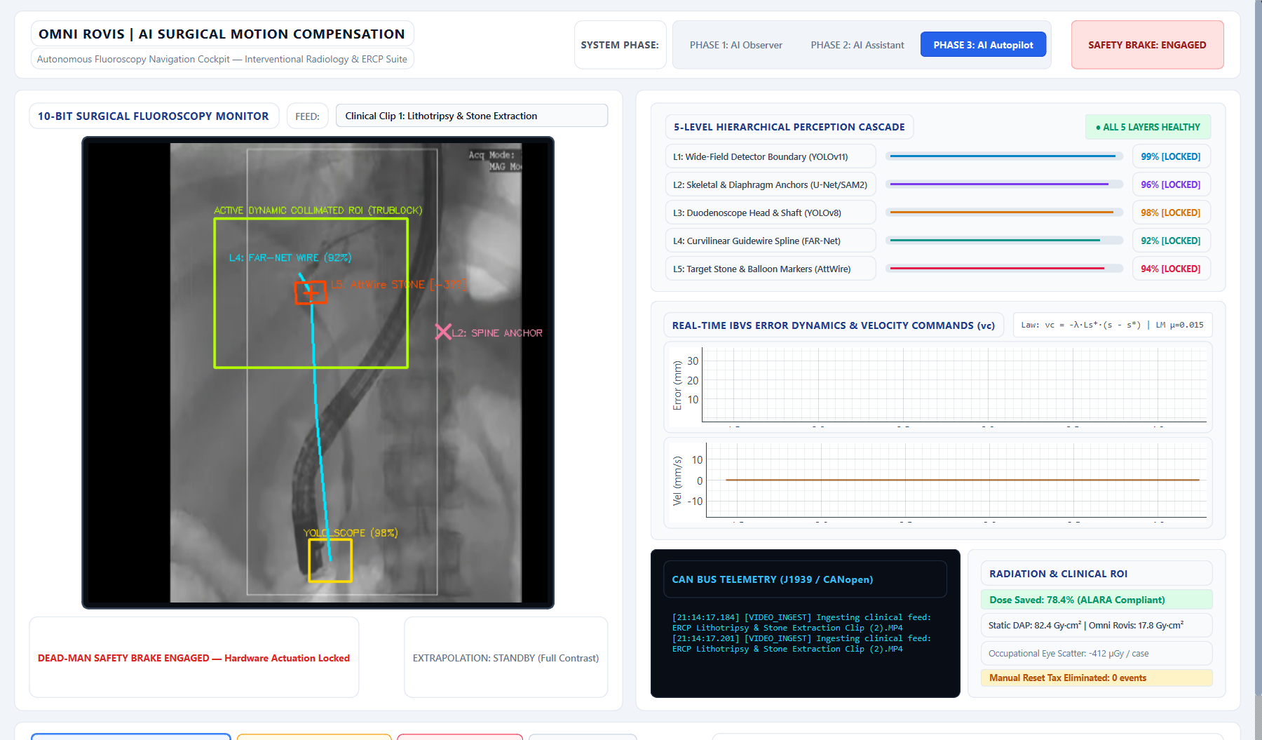
Complex Endoscopic Retrograde Cholangiopancreatography (ERCP)—such as difficult bile duct stones (>15 mm) or stricture cannulation—imposes severe physical, radiation, and cognitive burdens on surgical teams.
Published literature indicates complex therapeutic ERCP procedures often reach ~80+ Gy·cm² in Dose Area Product (DAP). Cumulative scatter elevates risks for cataracts and malignancies, while 15 kg lead aprons contribute to chronic surgeon orthopedic spine disease.
Under sedation, natural diaphragmatic excursions (14 BPM) and involuntary patient coughs displace the anatomical region of interest (ROI) by up to 79 mm. The surgeon must pause, drop instruments, and manually re-jog the C-arm or table, compounding fatigue.
Radiopaque iodine dye dissipates rapidly under continuous physiological bile flow. Once contrast clears, standard 2D tracking algorithms lose target visibility, forcing repeated contrast boluses or operating with degraded spatial certainty.
A unified multi-task perception backbone combined with Levenberg-Marquardt damped Image-Based Visual Servoing (IBVS) and patient-specific Bayesian motion calibration.
A single TensorRT feature extractor driving 5 lightweight task heads simultaneously, eliminating redundant compute and achieving sub-14ms closed-loop latency.
Locks onto global intensifier / Flat-Panel Detector (FPD) boundary to establish the physical image origin [270.0, 270.0].
Tracks rigid vertebral pedicle shadows and diaphragm curvature to form the spatial foundation for zero-contrast correlation.
Binds the distal duodenoscope articulation point at the ampulla, providing continuous relative mechanical grounding.
Extracts sub-pixel spline vectors tracing 0.035" hydrophilic guidewires through tortuous biliary duct geometry.
Computes 2D centroid and angular orientation theta of Dormia lithotripsy baskets, biliary stones, and stricture bifurcations.
Levenberg-Marquardt Damped Pseudoinverse: Calculates optimal instantaneous 3D velocity commands (\(v_c\)) for the motorized TruBlock collimator and C-arm. An adaptive gain law \(\lambda(e) = \lambda_{\text{base}} \cdot [0.80 + 0.20 \tanh(e/8.0)]\) enables rapid response to gross spasms (\(85\text{ mm/s}\)) while ensuring sub-millimeter damping near center without mechanical overshoot.
When contrast clears, a Bayesian Kalman Filter updates the target estimate using a patient-specific motion correlation matrix anchored to the L2 spine. The model recalibrates on every contrast pulse, maintaining an explicit confidence envelope.
Designed as a standalone beam-limiting add-on module requiring zero proprietary gantry write-access. Motorized lead shutters actively track the target, providing up to ~78% ALARA radiation shielding across peripheral tissue.
Redundant safety gating: (1) Hardware Dead-Man Foot Pedal clamps mechanical brakes in <12 ms; (2) AI Confidence Floor automatically freezes actuation if multi-head confidence drops below 70%, surfacing an immediate lost-lock alert.
Evaluated across three retrospective surgical video datasets comprising 2,654 ground-truth annotated frames.
Retrospective video evaluation of single-operator cholangioscopy mechanical lithotripsy for an impacted 16.5 mm bile duct stone. Under simulated 14 BPM respiration, the Dormia extraction basket oscillates across a 42 mm sweep.
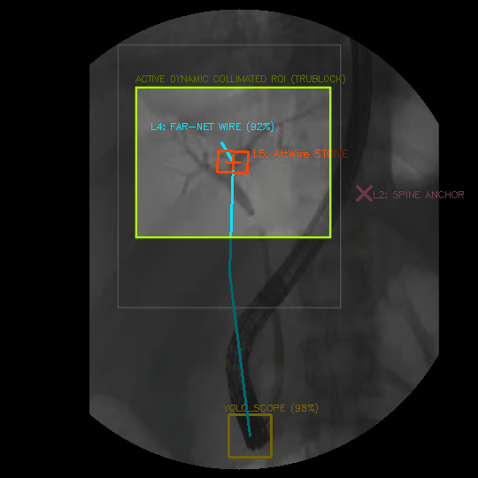 Pilot Clip 1: Lithotripsy basket and guidewire locked under simulated closed-loop IBVS.
Comparative evaluation against standard static ROI collimator behavior (fixed box baseline) during selective cannulation of the major duodenal papilla.
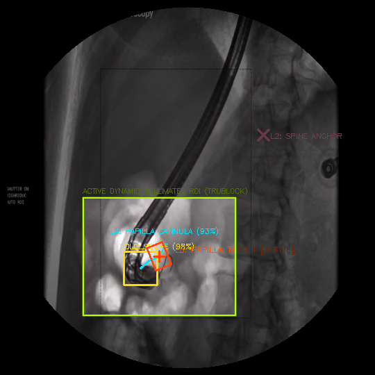 Pilot Clip 2: Omni Rovis dynamic collimation simulation vs. static background shutter.
Surgical suite workflow constraints during complex 71.52 mm proximal Common Bile Duct stricture dilation. Retrospective evaluation under severe noise and tool overlap.
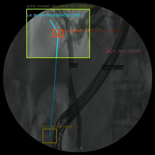 Pilot Clip 3: 71.5mm CBD stricture traversal with multi-task perception backbone.
Modeled radiation dose reductions (ALARA Tier 1) and operational time savings projected across standard high-volume interventional suites.
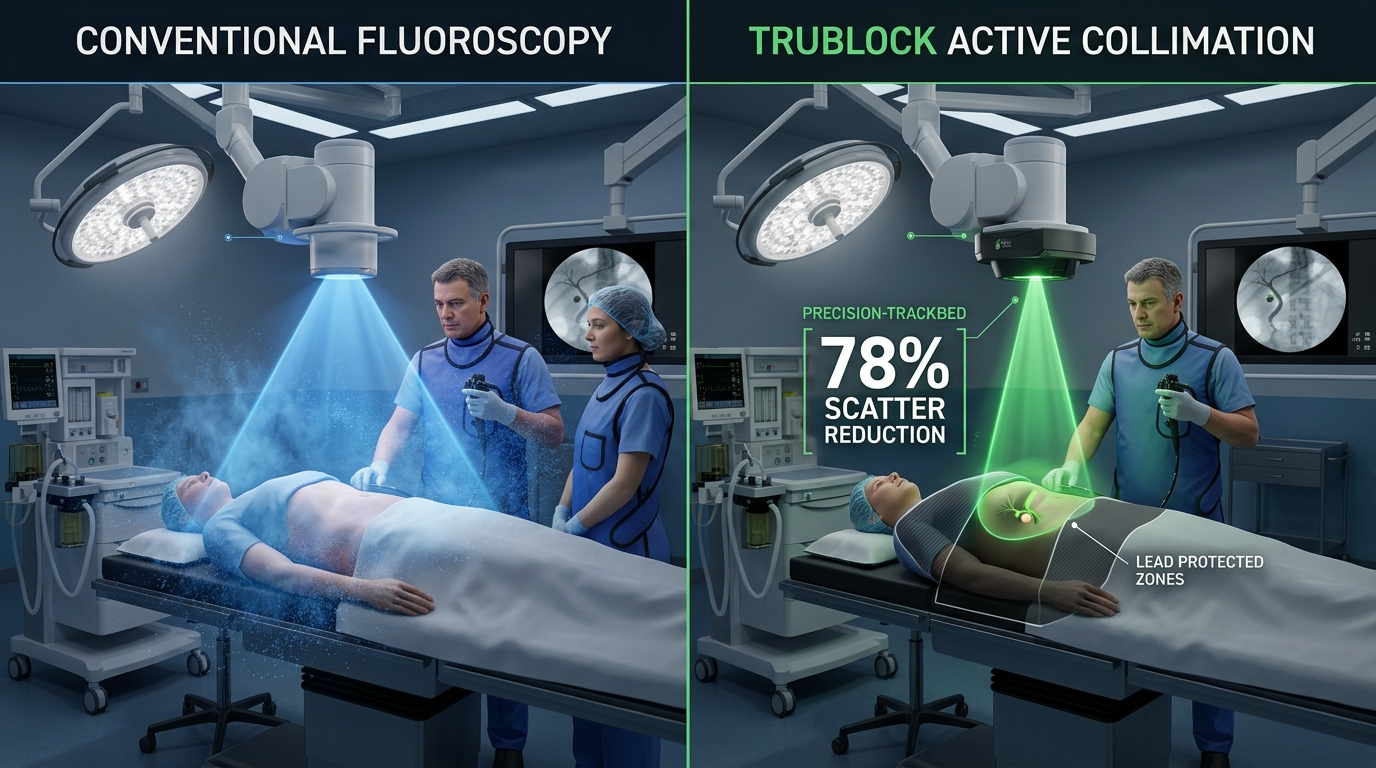
Wide conical beam floods the patient's entire abdomen, scattering heavily across the room and exposing the surgeon and nursing staff to high cumulative radiation.
Autonomous motorized lead shutters dynamically narrow the beam cone onto the biliary target, shielding peripheral organs and cutting occupational scatter by up to ~78%.
| Performance Metric | Conventional Fluoroscopy | Static ROI Shuttering Baseline | Omni Rovis (TruBlock Mode) |
|---|---|---|---|
| Mean DAP Exposure / Case | ~82.4 Gy·cm² (Literature) | ~42.1 Gy·cm² | ~17.8 Gy·cm² (Up to -78%)* |
| Occupational Eye Scatter (Staff) | ~528 µGy / case | ~295 µGy / case | ~116 µGy / case (-78%)* |
| Patient Motion Follow | None (Manual Jog) | Manual Box Drag | Autonomous Follow (<14ms latency) |
| Contrast Washout Handling | Zero (Visual Loss) | None | Bayesian Spine Motion Fusion |
| Manual Reset Interruption Rate | 12–18 pauses / case | 8–12 pauses / case | 0 pauses (Eliminated in Pilot) |
| Hardware Actuation Safety | Manual Foot Switch | Touchscreen UI | Dual: Dead-Man Pedal + Confidence Freeze |
Modeled across 450 ERCP cases/year via estimated 8.5 minute reduction in manual C-arm re-centering and fluoroscopy dead-time.
Designed to reduce reliance on heavy lead shielding during prolonged procedures, supporting surgeon orthopedic health.
Architecture designed to comply with IEC 62304 Class C ↗ medical software life-cycle and ALARA radiation minimization guidelines.
A pragmatic commercial strategy starting with a standalone retrofit collimator, expanding into OEM gantry navigation partnerships.
Instead of attempting to command a 300 kg hospital machine, TruBlock leaves the C-arm completely frozen and drives 4 high-speed lead shutter blades to steer the X-ray beam directly.
Taps into the C-arm monitor's auxiliary Video-Out / DVI port. 100% passive and read-only. Zero modifications to proprietary Siemens, GE, or Philips firmware.
Unified multi-task TensorRT vision backbone tracks the duodenoscope, guidewire, and moving biliary stone, calculating the exact spatial drift vector.
Four NEMA 14 stepper motors slide 2.5 mm lead blades across miniature linear rails. The 300 kg machine stays frozen; the beam follows the stone!
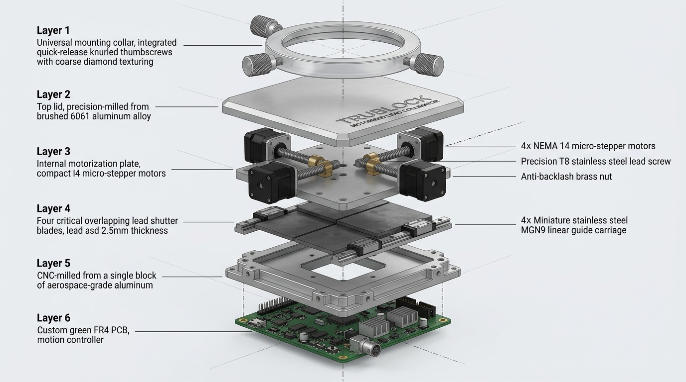
Quick-release universal mounting collar, brushed 6061-T6 aluminum housing, 4x NEMA 14 micro-steppers with T8 lead screws, 4x overlapping 2.5mm lead blades on MGN9 rails, and custom STM32 motion controller PCB.
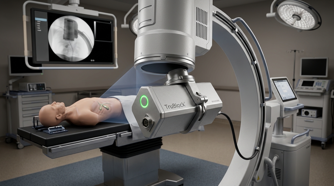
Clamps securely onto standard 80–140mm tube ports in under 60 seconds without tools. Compact 8 kg module footprint with single umbilical cable routing to the mobile workstation console.
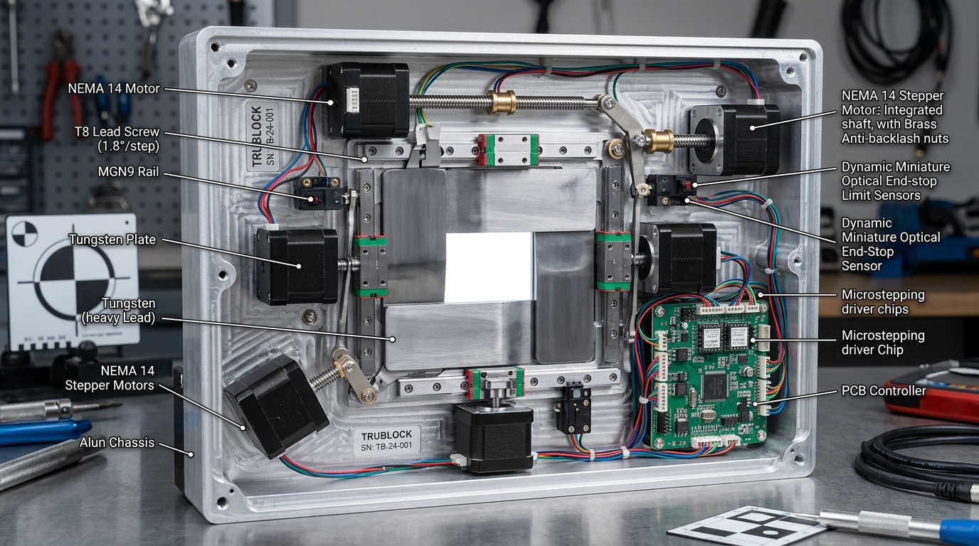
Suspended on dual stainless steel MGN9 linear guide carriages and driven by TMC2209 silent microstepping drivers. A 5 mm respiration stroke is traversed in <35 milliseconds with zero audible clicking in the sterile operating field.
Adopting the programmable dynamic respiratory motion phantom methodology established by leading robotic radiotherapy systems (e.g. Accuray CyberKnife) to prove sub-millimeter tracking accuracy against known encoder ground truth.
Trained and benchmarked unified multi-task TensorRT vision backbone across 2,654 ground-truth surgical frames (<14ms latency, 0.24mm pilot error).
View 3 Ground-Truth JSONs ↗Closed-loop visual servoing validation on a programmable motorized respiratory stage driven by recorded asymmetric human breathing traces.
AAPM TG-76 Protocol (PDF) ↗Benchtop integration of autonomous motorized lead shutter retrofit module (Class II 510k Beam-Limiting Device regulatory pathway).
FDA 21 CFR 892.1610 ↗In vitro phantom evaluation on academic interventional radiology research units with open API control access.
IEC 62304 Medical Standard ↗Commercial software licensing and CANopen integration with major global C-arm manufacturers (Siemens, GE, Philips).
OmniIDE Research Group ↗Omni Rovis is developed by the AI research and robotics division at OmniIDE, based in Bengaluru, India. We are actively engaging with interventional gastroenterologists, hospital research institutions, and MedTech investors.
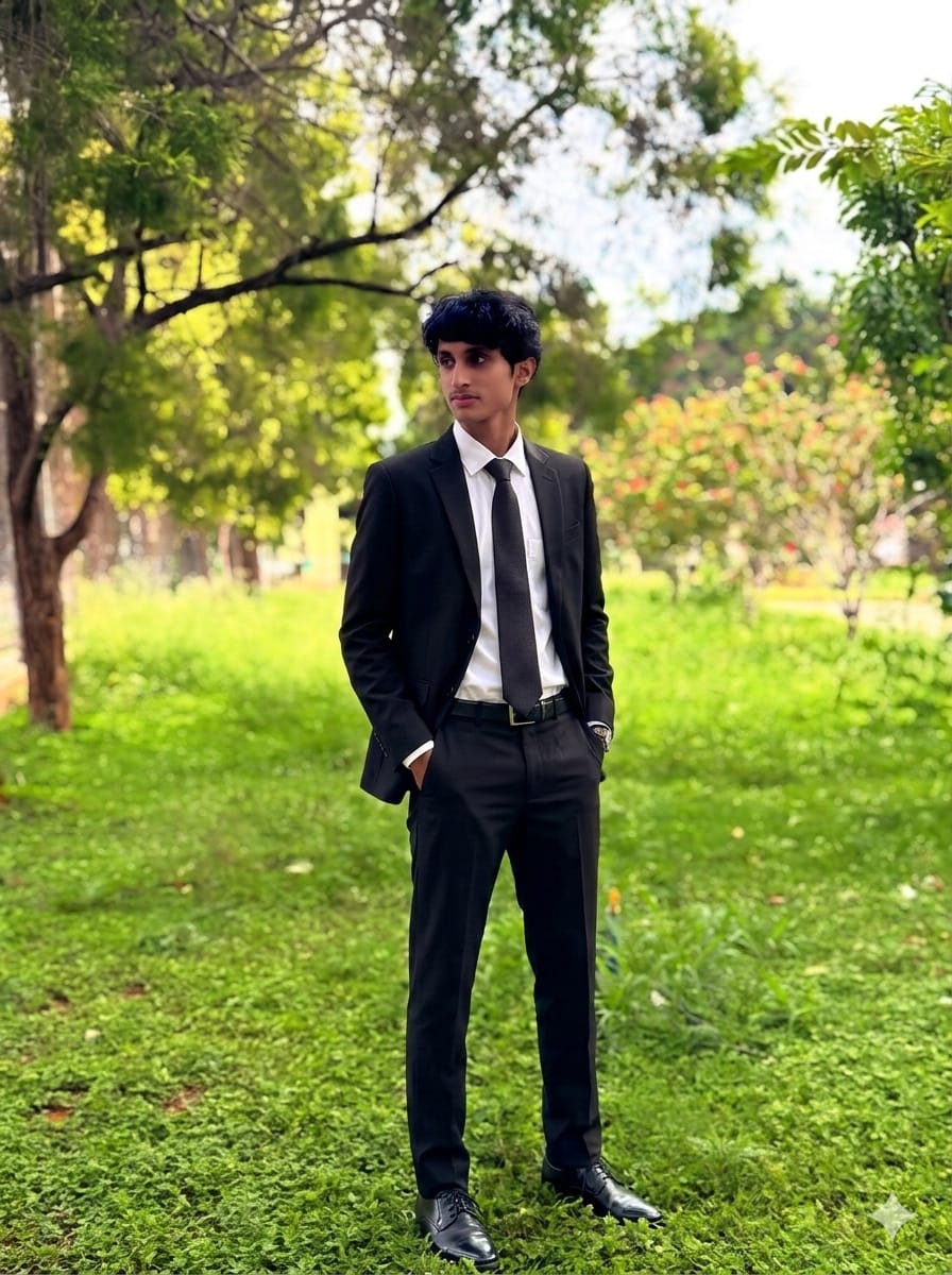
Founder & CEO of OmniIDE and Omni Rovis. Leading AI architecture, real-time visual servoing control systems, and algorithmic medical motion compensation infrastructure.

AI research and engineering organization building autonomous intelligence systems and medical robotics software. Registered enterprise (UDYAM-KR-03-0681404).
Receive our comprehensive technical whitepaper, IBVS mathematical derivation dossier, retrospective video benchmarks, and regulatory roadmap directly from our founding team.